Fig. 5.
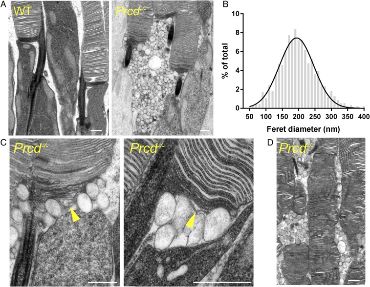
Accumulation of extracellular vesicles at the base of photoreceptor outer segments in Prcd−/− mice. (A) Electron micrographs of WT and Prcd−/− rod photoreceptors. Note that contrasting membranes with tannic acid yields darker staining of newly forming discs than mature, enclosed discs. (B) Gaussian histogram of Feret’s maximum diameters for the extracellular vesicles accumulating next to the outer segments of Prcd−/− mice. (C) High-magnification electron micrographs of the site of disc morphogenesis in Prcd−/− mice. Arrowheads indicate newly forming discs, which are bulging. (D) Electron micrograph illustrating uneven outer segment diameter of Prcd−/− rods. All mice were 2-mo old (Scale bars in all panels, 500 nm).

