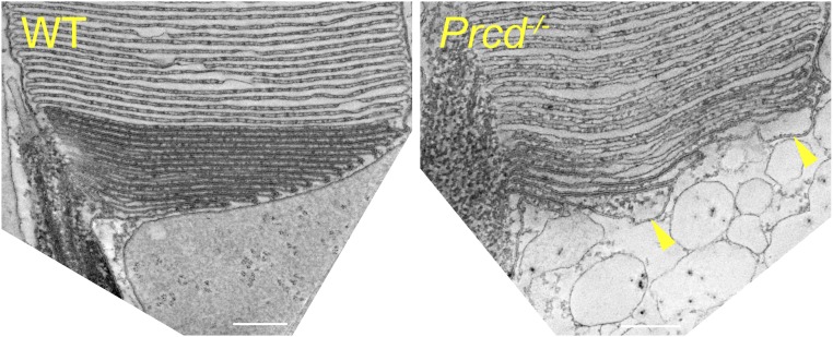
Fig. 7.
Three-dimensional electron tomography of WT and Prcd−/− rods at the site of disc morphogenesis. An image of a single, 1-nm z-section from ∼250-nm-thick electron tomograms of WT and Prcd−/− rods taken at the site of disc morphogenesis. The entire tomograms are shown in Movies S1 and S2. Arrowheads indicate two prominent bulges at the edges of newly forming discs of the Prcd−/− rod. Mice were 2-mo old (Scale bars, 200 nm).

