Fig. 4.
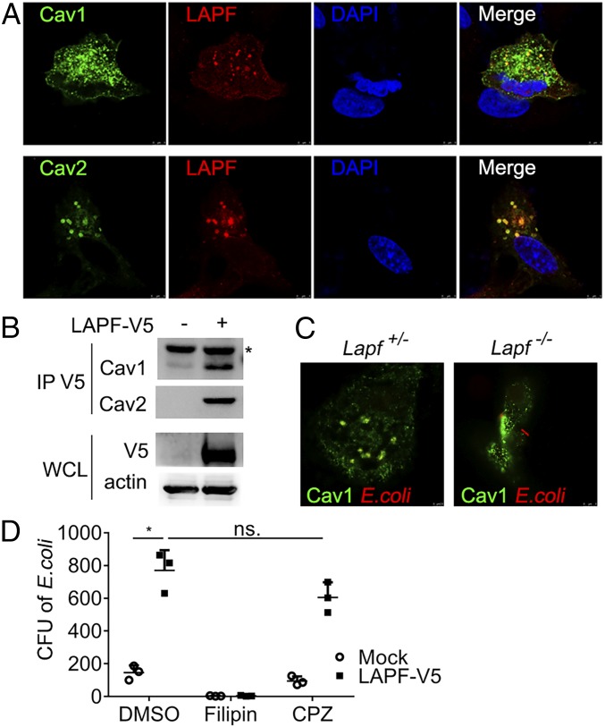
LAPF interacts with Cav1/2 and promotes caveolae-dependent endocytosis. (A) Confocal microscopy of the immunofluorescence staining of overexpressed LAPF and Cav1/2 in A549 cells. (Magnification, 252×.) (B) Immunoblot analysis of anti-V5 immunoprecipitated lysates of 293T cells. (C) Confocal microscopy of the immunofluorescence staining of Cav1 and E. coli in Lapf+/− and Lapf−/− macrophages. (Magnification, 252×.) (D) CFU assay after E. coli infection in macrophages pretreated with Filipin (3 μM) or CPZ (10 μM). Data are presented as mean ± SD of three independent experiments (D), or shown for one representative experiment from three independent experiments with similar results (A–C). *P < 0.05. ns., no significant differences.

