
Figure 4.
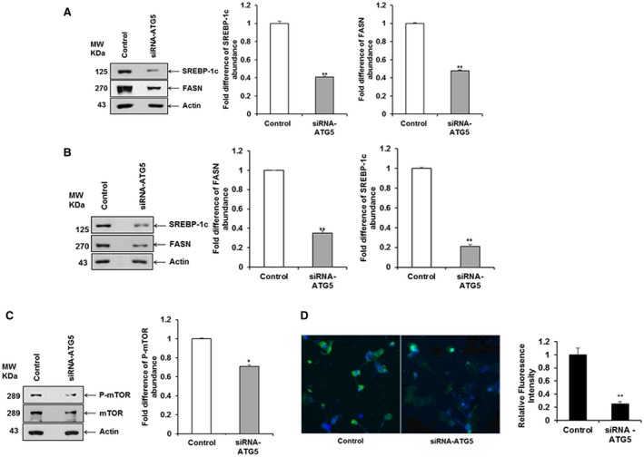
Knockdown of ATG5 inhibits SREBP‐1c and FASN in hepatocytes. (A) Huh7.5 cells were transfected with siRNA to ATG5 (siRNA–ATG5) or siRNA negative control, and cell lysates were prepared after 48 hour post‐transfection. SREBP‐1c and FASN were detected by western blot analysis using specific antibodies. The blot was reprobed with antibody to actin for loading control. (B) Huh7.5 cells harboring the HCV genome‐length replicon cells were transfected for 48 hours with siRNA–ATG5 or siRNA negative control, and cell lysates were prepared. SREBP‐1c and FASN were down‐regulated in siRNA–ATG5‐transfected hepatocytes. (C) A significant down‐regulation of p‐mTOR expression level in Huh7.5 cells transfected with siRNA–ATG5 compared to that of the control cells was observed after normalization with actin. A densitometry scan is shown on the right in all panels. (D) Hepatocytes were transfected for 48 hours with siRNA–ATG5 or siRNA negative control, and cells were stained 48 hours after transfection. Confocal microscopy examination for LDs visualized by LDs (BODIPY, green) and nucleus (DAPI, blue) in merged image panels (20×). A representative image is shown. Quantitation of the BODIPY staining is shown in the bar graph on the right. Statistical significance was analyzed using the two‐tailed Student t test; *P < 0.05, **P < 0.01. Abbreviation: BODIPY, boron‐dipyrromethene.