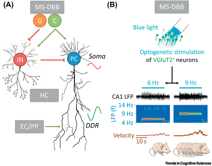
Figure I. Mechanistic basis of theta oscillations.
(A) Classic theta model (adapted from [17]). See Box 2 for explanations. Note that many extensions have been suggested to this model. C, cholinergic; DDR, distal dendritic region; EC/PP, entorhinal cortex and perforant path; G, glutamatergic; HC, hippocampus; IN, interneuron; MS-DBB, medial septum and diagonal band of Broca; PC, pyramidal cell. (B) MS-DBB VGluT2+ neurons as a key player in regulating locomotion. Increasing optogenetic stimulation (blue light) frequency of these neurons (from 6 to 9Hz) leads to higher theta oscillation frequency in CA1 and is followed by increased velocity of the rodent (adapted from [34]).

