Figure 5.
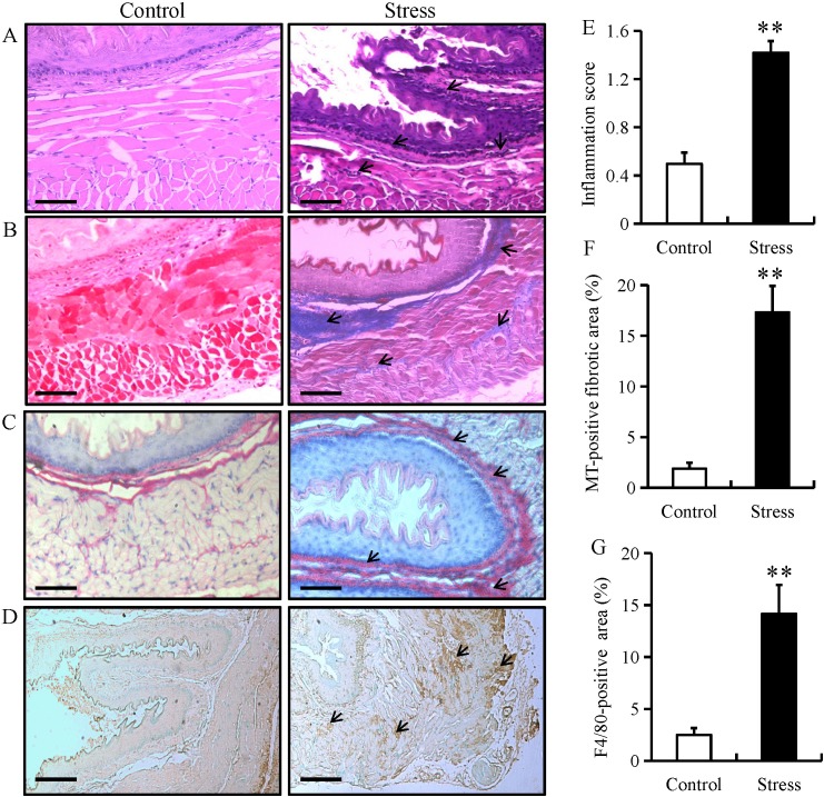
Chronic restraint stress induces esophageal fibrosis in mice. Esophageal tissue from mice in the chronic restraint stress and control groups were analyzed by (A) H&E, (B) MT and (C) Sirus red staining. (magnification, ×200; scale bar, 50 µm). (D) Immunostaining for F4/80 in esophageal tissue from mice in the chronic restraint stress group and control group (magnification, ×200; scale bar, 50 µm). (E) Histology inflammation score. (F) Quantification of the MT-positive areas. (G) Quantification of the F4/80-positive areas. Data presented as the mean ± standard deviation (n=15). **P<0.001 vs. control. H&E, hematoxylin and eosin; MT, Masson's trichrome.

