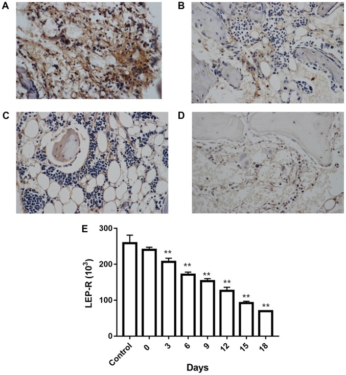
Figure 3.
Immunohistochemical analysis of LEP-R levels in the bone marrow of: (A) Control mice; and AA model mice at (B) 6 days, (C) 12 days and (D) 18 days after infusion of lymph node cells. (E) Changes in LEP-R levels in the AA model mice. **P<0.01 vs. control group. LEP-R, leptin receptor; AA, aplastic anemia.

