
Figure 4.
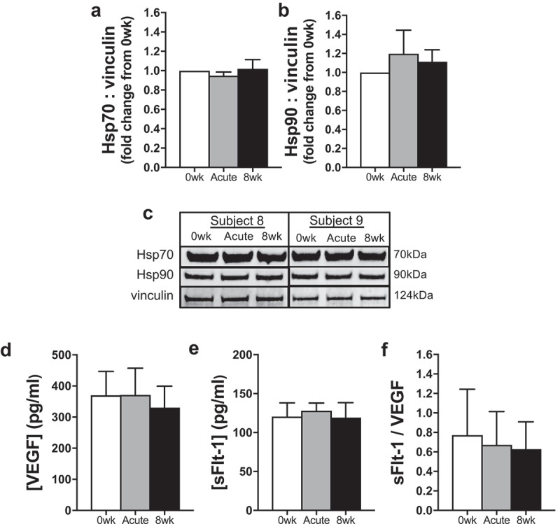
(a–b) Protein abundance of heat shock protein (Hsp) 70 (a) and Hsp90 (b) in endothelial cells exposed to sera from human subjects collected before (0 wk) and 1 h after (Acute) the first hot water immersion session and following 8 wk of heat therapy. Data were normalized to a vinculin loading control and presented as mean±SE fold changes from 0 wk. (c) Representative Western blot images are provided below. (d–f) Serum concentrations of vascular endothelial growth factor (VEGF) (d) and soluble VEGF receptor (sFlt)-1 (e), and the ratio of sFlt-1/VEGF, indicative of bioavailable VEGF, across time points into heat therapy (f). Data are mean±SE No significant differences were observed.