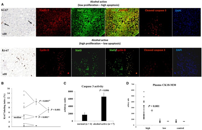
Figure 4.

Proliferation–apoptosis balance in early‐stage ALD in actively drinking patients. (A) Representative immunohistochemistry of Ki‐67, immunofluorescence of cyclin D, Stat3, and cleaved caspase 3 showing a low proliferation, high apoptosis (upper panel), and high proliferation, low apoptosis group (lower panel). Stat3 was sequestered in the cytosol of low proliferators, but co‐localized with cyclin D in the nucleus of high proliferators. (B) Quantification of Ki‐67 expression confirming the presence of a high, intermediate, and low proliferation group. P values refer to the low (*) and intermediate ($) proliferation group. (C) Caspase 3 activity in liver lysates of controls and patients with high cleaved caspase 3 nuclear expression confirming increased apoptosis. (D) Increased blood levels of the apoptotic cytokeratin 18 fragments M30 confirming the presence of a high and low apoptosis group. P value compares to the low apoptosis group and to controls. Abbreviation: DAPI, 4′,6‐diamidino‐2‐phénylindole.
