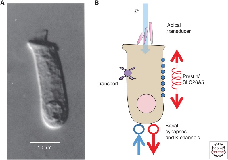
Figure 1.
The elements of the mammalian outer hair cell (OHC). (A) An isolated OHC from turn 2 (∼5–10 kHz region) of the guinea pig cochlea. The apical stereocilia are apparent at the apical surface. (B) Schematic OHC showing the location of prestin/SLC26A5 down the basolateral surface, generating longitudinal forces. Transport to regulate pHi is presumed to be collocated as a parallel property of prestin. K+ ions from scala media enter through the mechanoelectric transducer channels at the apex and exit through the basally located K+ channels. Both afferent (red) and efferent (blue) terminals are located at the base of the cell.

