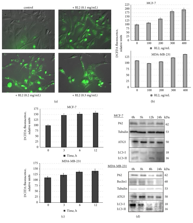
Figure 2.
Analysis of ROS production and changes in autophagy-related proteins. (a) Representative fluorescence microscopy images of ROS production (green) in RL2-treated MDA-MB-231 cells. (b), (c) Dependence of ROS production on RL2 concentration and time of incubation. For a kinetic study of ROS production, cells were treated with RL2 (300 μg/mL). (d) The representative Western Blot of MCF-7 and MDA-MB-231 cells treated with RL2 (300 μg/mL) is showing a level of autophagy-related proteins.

