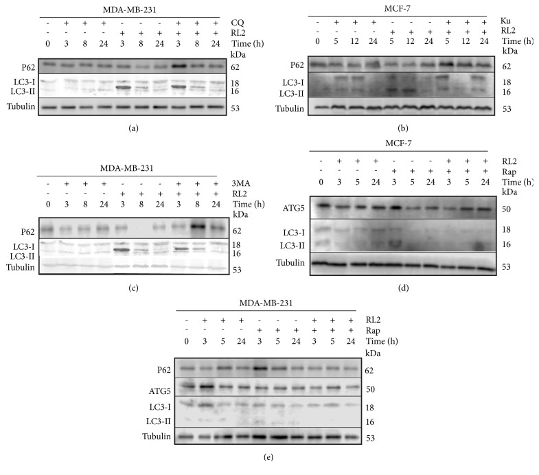
Figure 5.
Changes in the cellular proteins after their incubation with indicated compounds. Representative Western Blots showing changes in autophagy-related proteins. Whole cell lysates were prepared for analysis using tubulin as a loading control. (a), (c), (e) Western Blots with MDA-MB-231 cell lysates. Cells were treated with RL2 (0.3 mg/mL), CQ (10 μM), 3MA (10 mM), and Rap (10 μM) for various time points (0–24h). (b), (d) Western Blots with MCF-7 cell lysates. Cells were treated with RL2 (0.3 mg/mL), Ku (30 μM), and Rap (10 μM) for the indicated time points (0–24h).

