Figure 2.

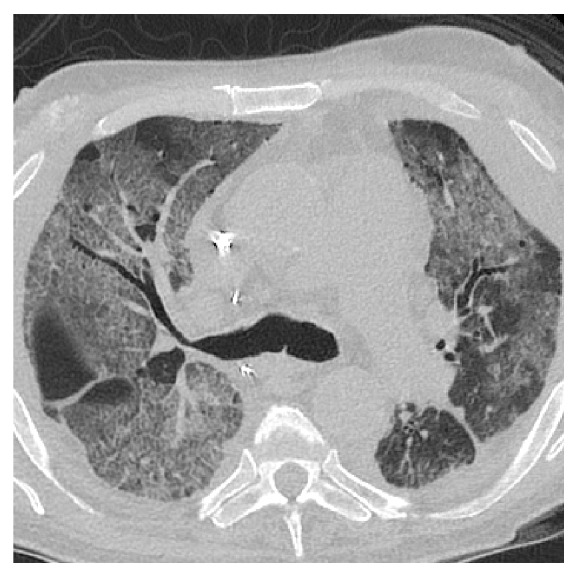
Transverse chest computed tomography demonstrating widespread peribronchovascular ground-glass opacities throughout both lung fields.

Transverse chest computed tomography demonstrating widespread peribronchovascular ground-glass opacities throughout both lung fields.