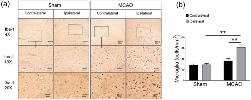
Figure 1.

Microglial cells were increased after ischemic stroke. (a) Representative images showed that microglia increased in the ipsilateral side after MCAO. (b) Semi-quantification analysis of the number of Iba-1 positive cells. Data were presented as mean± S.E.M., *p < 0.05, **p < 0.01. Five random fields were counted for each sample, n = 5 per group.
