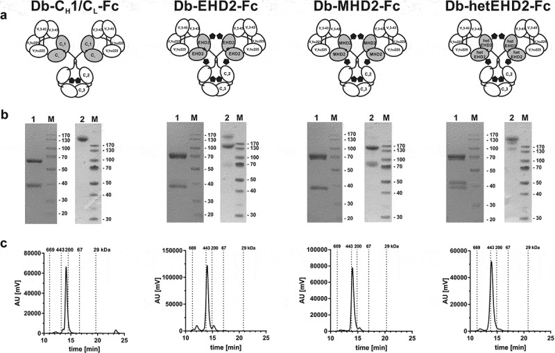
Figure 2.

Biochemical characterization of Db3-43xhu225-Ig molecules. (a) Schematic illustration of Db3-43xhu225-Ig molecules using CH1/CL, EHD2, MHD2, or hetEHD2 as dimerization module. (b) SDS-PAGE analysis of Db3-43xhu225-Ig molecules under reducing (1) or non-reducing (2) conditions (4–12% PAA gradient). Proteins were stained with Coomassie blue. (c) Size-exclusion chromatography of Db3-43xhu225-Ig molecules by HPLC using a Tosoh TSKgelSuperSW column.
