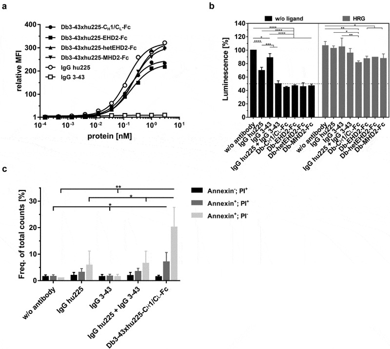
Figure 4.

Binding and bioactivity of bispecific Db3-43xhu225-Ig molecules on FaDu cells. (a) Binding of different antibodies to FaDu cells was analyzed by flow cytometry. Bound antibodies were detected using a PE-labeled anti-human Fc secondary antibody. Mean ± SD, n = 3. (b) Proliferation assay of different bispecific Db3-43xhu225-Ig molecules (50 nM) using FaDu cells in starvation medium after incubation of 7 days. Cells were kept either unstimulated (w/o ligand) or stimulated with heregulin (HRG). Parental antibodies were included as control (single treatment: 50 nM; combination: 50 nM each). Cell viability was measured using CellTiter-Glo 2.0. Mean ± SD, n = 3. (c) Flow cytometry analysis of annexinV/PI staining using FaDu cells. Bispecific Db3-43xhu225-CH1/CL-Fc molecule (50 nM), or parental antibodies (single treatment: 50 nM; combination: 50 nM each) were incubated with cells in complete medium for 24 h. Mean ± SD, n = 3. *p < .05; **p < .01; ***p < .001; ****p < .0001.
