
Figure 2.
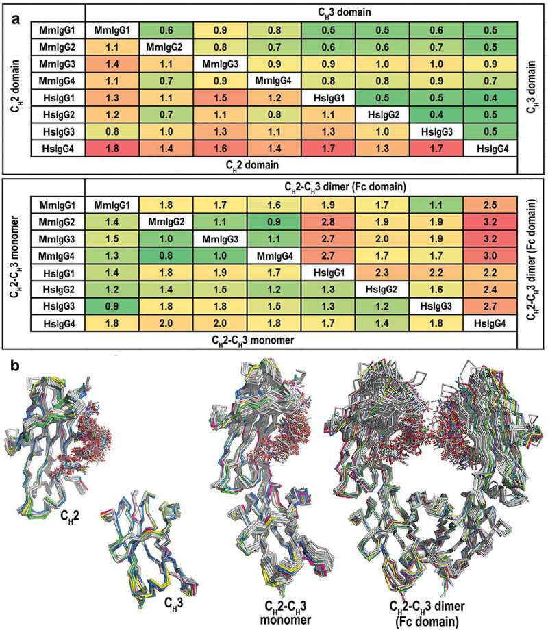
Comparison of the overall structures of the Rhesus macaque and human IgG1-4 Fc. (a) Average RMSD values for main chain atoms for pairwise comparisons of CH2, CH3, CH2-CH3 monomers and CH2-CH3 dimers (Fc domain). The structures of the Fcs of human IgG1-4 used in the alignments include: IgG1, PDB codes: 3AVE, 4DZ8, 4W4N, 1H3Y and 1H3V; IgG2, PDB codes: 4HAF, 4HAG; IgG3, PDB code: 5W38 and IgG4, PDB codes: 4C54, 4C55, 5LG1. (b) Structural alignment of CH2, CH3, CH2-CH3 monomers, and CH2-CH3 dimers. MmFcs are colored in green for IgG1, pink for IgG2, blue for IgG3 and yellow for IgG4. Human Fcs are colored in grey. CH2-CH3 dimers are aligned by superimposing the CH3 domains.