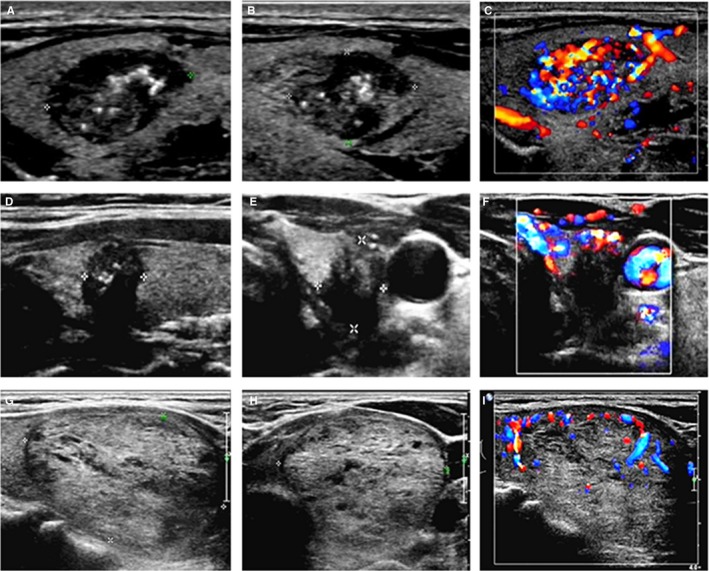
Figure 4.

Ultrasonic images of different kinds of thyroid nodules. (A‐C) were ultrasound images of a case of MTC. Male patient, 49 years old. (A) Lesion was solid, markedly hypoechoic, well‐defined, with microcalcification. (B) A/T < 1. C. Enhanced blood flow. (D‐F) were ultrasound images of a case of PTC. Female patient, 47 years old. (D) Lesion was solid, markedly hypoechoic, with microcalcification, ill‐define. (E) A/T ≥ 1. (F) Absent of blood flow. (G‐I) were ultrasound images of a case of benign nodules. Female patient, 63 years old. (G) Lesion was almost solid, isoechoic, well‐defined, with none of calcifications. (H) A/T < 1. (I) Few blood flow. MTC, medullary thyroid carcinoma; PTC, papillary thyroid carcinoma; A/T ≥ 1, the shape of nodule is taller‐than‐wide; A/T < 1, the shape of nodule is wider‐than‐tall
