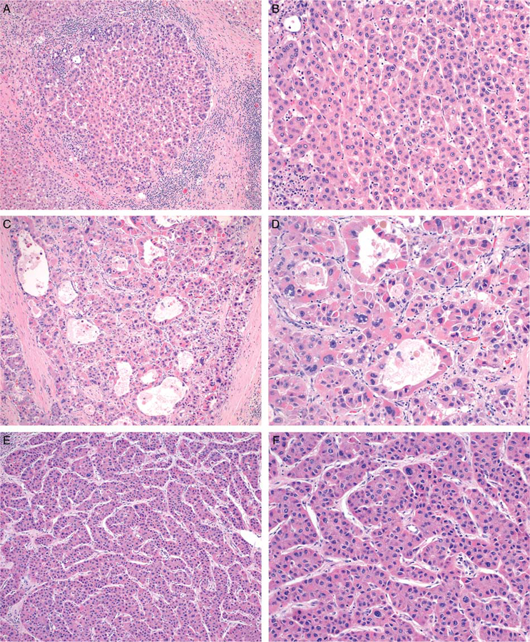
FIGURE 2.
The degree of nuclear pleomorphism falls into two categories. Tumors without significant pleomorphism (A, B) contain relatively uniform nuclei. Those with significant pleomorphism show nuclear size variation greater than 2× in at least two foci within a single 20× field. Some cases show diffuse nuclear pleomorphism (C, D) while this feature is only focally present in others (E, F) (H&E).

