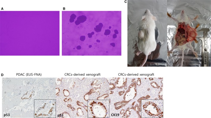
Figure 4.

Tumorigenesis in vitro and in vivo. (A, B), In vitro tumorigenesis, Soft agar colony formation assay. (A) Negative control, (B) tumor cell formation at day 15; (C‐E) In vivo tumorigenesis and representative histology of xenografts. Hematoxylin and eosin staining showed that the implanted patient‐derived CRCs had a ductal structure. (C) In vivo tumorigenesis, 5‐week‐old male nonobese diabetic/severe combined immunodeficiency mice; (D) CRC‐derived xenograft tissue demonstrated homology for p53 expression compared with matched primary biopsy cancer tissue. The Ductal epithelial marker CK19 is positive in CRC‐derived xenograft by IHC stain
