
Figure 9.
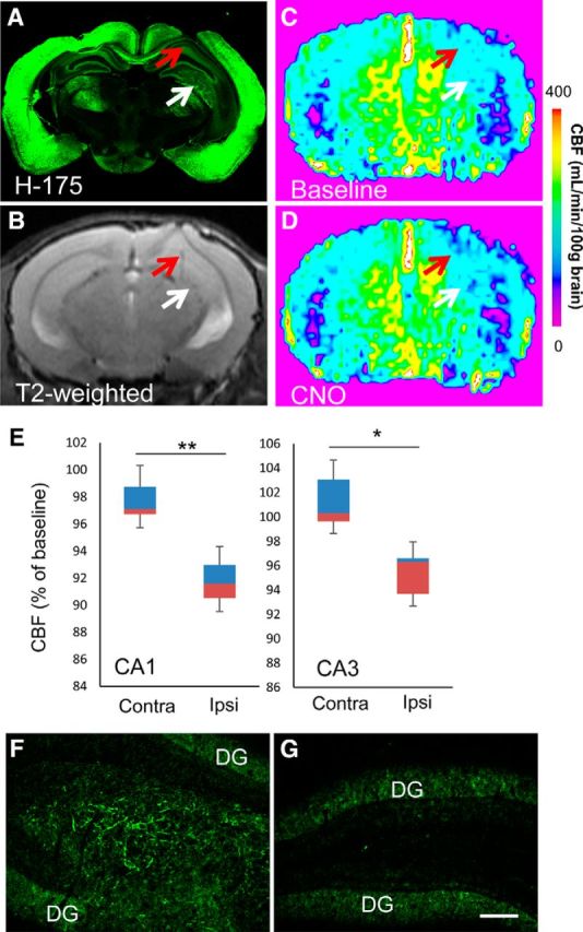
In vivo imaging assessments of the functionality of hM4Di-iPSC-derived neuronal grafts. A–D, Representative postmortem microscopic (A) and antemortem T2-weighted (B) and ASL (C, D) MRI images of an adult mouse brain at 40 d after implantation of hM4Di-iPSC-derived grafts. A low-power photomicrograph illustrates immunolabeling of endogenous and graft-derived M4 mAChRs with H-175 (A). Red and white arrows indicate the ipsilateral hippocampal CA1 sector and hilus, respectively. This mouse underwent repeated ASL-MRI at 40, 46, and 52 d after implantation. Scale bar indicates CBF measures in C and D. E, Quantitative assays of CNO-induced CBF changes in hippocampal CA1 and CA3 sectors. Data are displayed in boxplots. The bottom and top of rectangles indicate the first and third quartiles, respectively. A horizontal line inside the rectangles shows the median and the “whiskers” above and below the rectangles express the maximum and minimum values, respectively. Quantitative analysis of these three assays demonstrated that CBF was decreased significantly in the implantation site (hilus/CA3), as well as the ipsilateral CA1 sector, in response to CNO (n = 3, t = 15.808 and t = 4.635 for CA1 and hilus/CA3, respectively, *p < 0.05, **p < 0.01, by paired t test, versus the corresponding contralateral regions). The brain of this mouse was collected after the last MRI scan for postmortem analysis. F, G, H-175 immunoreactivity in the hippocampal hilus (F) implanted with hM4Di-iPSC-derived cells, in sharp contrast with the absence of detectable signals in the contralateral hilar region (G). Immunofluorescence signals in the dentate gyrus (DG) were primarily derived from expression of endogenous M4 mAChR. Ipsi, Ipsilateral; Contra, contralateral. Scale bar, 100 μm (F, G).