Figure 7.
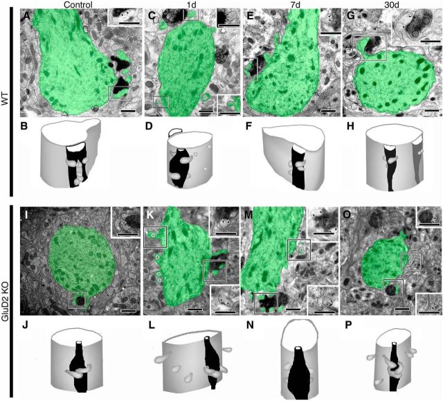
Electron micrographs and 3D reconstructed images of CF synapses on proximal dendrites in sham-operated control (A, B, I, J) and PF-transected mice on postlesion days 1 (C, D, K, L), 7 (E, F, M, N), and 30 (G, H, O, P) in wild-type (A–H) and GluD2-KO (I–P) genotypes. In electron micrographs, PC dendrites/spines are pseudocolored in green, whereas CFs are labeled by dark precipitates for VGluT2. Insets, Enlarged images of PF–PC synapses in boxed regions. Pairs of arrowheads and asterisks indicate the PSD and free spines, respectively. Note the emergence of free spines at proximal dendrites on postlesion day 1 in wild-type mice (C, D) and their frequent occurrence on all postlesion days in GluD2-KO mice (K–P). Scale bars, 500 nm.

