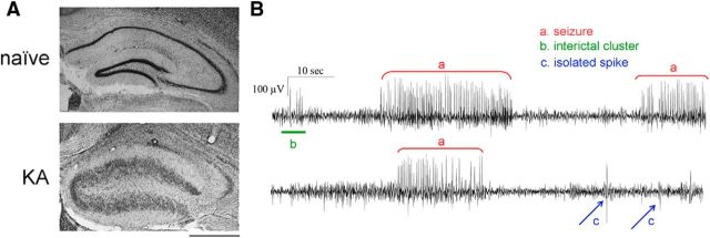
Figure 1.
Histopathological changes and EEG seizures in the KA model of MTLE. A, Nissl staining in coronal sections from a naive mouse (top) and from a mouse killed 21 d after intrahippocampal KA (bottom). In the epileptic hippocampus, note the typical granule cell dispersion and extensive neuronal loss in CA1 and CA3. Scale bar, 500 μm. B, Representative EEG recordings displaying activity in the hippocampus during the chronic phase of epilepsy. Three types of epileptiform events are distinguished: seizures (spike clusters lasting for >4 s; a, red), interictal clusters (spike clusters lasting <4 s; b, green), and isolated spikes (c, blue).

