Figure 2.
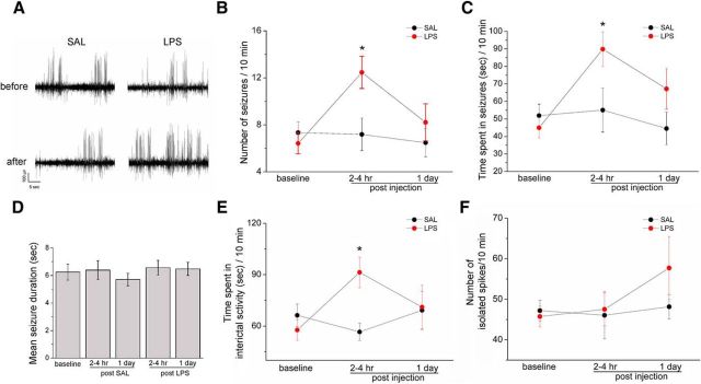
Impact of LPS challenge on the epileptic brain. A, Representative intrahippocampal EEG recordings from chronically epileptic mice, before and 2–4 h after saline/LPS administration. LPS clearly increases SRS frequency. B, Number of EEG seizures per 10 min of recording in epileptic mice, before and after saline (black circles) or LPS (red circles). Seizure frequency is significantly enhanced by LPS (two-way repeated-measures ANOVA followed by Holm–Sidak test, p < 0.05). C, Total time spent in ictal activity per 10 min of recording. Epileptic animals treated with LPS significantly spent more time in ictal activity than saline-injected mice (two-way repeated-measures ANOVA followed by Holm–Sidak test, p < 0.05). D, Mean duration of EEG seizures in saline- and LPS-treated mice (one way ANOVA; p = 0.7). E, Total time spent in interictal activity per 10 min of recording. Epileptic animals treated with LPS (red circles) significantly spent more time in interictal activity than saline-injected mice (black circle) 2–4 h following the systemic challenge (two-way repeated-measures ANOVA followed by Holm–Sidak test, p < 0.05). F, Number of isolated spikes per 10 min of recordings. No difference was found between LPS and saline-injected epileptic mice (two-way repeated-measures ANOVA followed by Holm–Sidak test, p = 0.5). Data are ± SEM. *p < 0.05.

