
Figure 6.
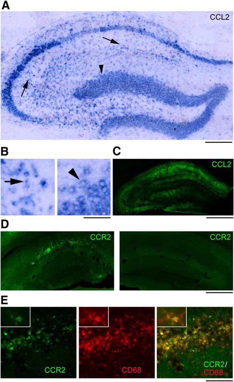
Cellular localization of CCL2 and CCR2 in the sclerotic hippocampus after LPS. A–C, CCL2 mRNA (A, B) and protein (C) staining on representative sections from the dorsal hippocampus of an epileptic mouse, 4 h after LPS. CCL2 staining is present in pyramidal neurons, dispersed granule cells of the dentate gyrus (A, B, arrowhead) and several small, likely glial cells of stratum oriens, stratum radiatum (A, B, arrows), stratum lacunosum moleculare, and hilus. Details of labeled cells are shown in B. Left, CA1 stratum radiatum. Right, Dentate granule cells. Scale bars: A, 200 μm; B, 40 μm; C, 600 μm. D, Immunostainings of CCR2 on representative sections from the epileptic (left) and contralateral (right) hippocampus, 4 h after LPS. CCR2 staining is mainly restricted to the CA1 subfield and dentate gyrus of the epileptic hippocampus and is absent from the contralateral side. Scale bar, 600 μm. E, Immunostainings of CCR2 (green), CD68 (red), and their colocalization (yellow) on a representative section from the CA1 subfield of an epileptic mouse, 4 h after LPS. Expression of CCR2 in activated microglia/macrophages, as indicated by CCR2/CD68 colocalization (yellow), is evident in high-magnification figures shown in insets. Scale bar: 150 μm; insets, 30 μm.