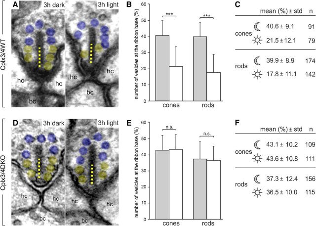
Figure 10.
Quantification of synaptic vesicles associated with the ribbon base in Cplx3/4 WT and Cplx3/4 DKO cone and rod photoreceptors. A, D, Electron micrographs of cone photoreceptor ribbons from Cplx3/4 WT (A) and Cplx3/4 DKO (D) retinas fixed after 3 h of dark adaptation and 3 h of light adaptation. Ribbon-associated vesicles within the basal 100 nm (dotted line) of the ribbon are highlighted in yellow, the remaining ribbon-associated vesicles in blue. B, E, Percentage (mean ± SD) of vesicles at the ribbon base in cone and rod photoreceptor ribbons from Cplx3/4 WT (B) and Cplx3/4 DKO (E) retinas fixed after 3 h of dark (gray bars) adaptation and 3 h of light (white bars) adaptation. ***p < 0.001, t test. C, F, Summary of the quantitative data. n = number of quantified ribbons; hc, horizontal cell; bc, bipolar cell. Scale bar in A applies to A and D, 100 nm.

