Figure 9.
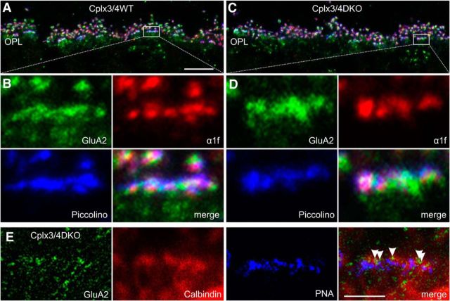
Molecular anatomy of cone photoreceptor ribbon synaptic sites in Cplx3/4 WT and Cplx3/4 DKO mouse retinas. A, C, Confocal laser scanning image of the outer plexiform layer (OPL) of Cplx3/4 WT (A) and Cplx3/4 DKO (C) retinas triple labeled with antibodies against GluA2 (green), L-type Ca2+ channel subunit α1f (red), and Piccolino (blue). B, D, High-power views of the boxed areas in A and C. E, High-power view of a Cplx3/4 DKO cone photoreceptor terminal triple labeled with antibodies against GluA2 (green), Calbindin (red), and peanut agglutinin (PNA, blue). The merge displays an overlay of a confocal (Calbindin, PNA) and a STED (GluA2) image. Arrowheads point to single GluA2-positive clusters at the cone photoreceptor terminal. Scale bar in A applies to A and C, 10 μm; scale bar in E, 2 μm.

