Figure 5.
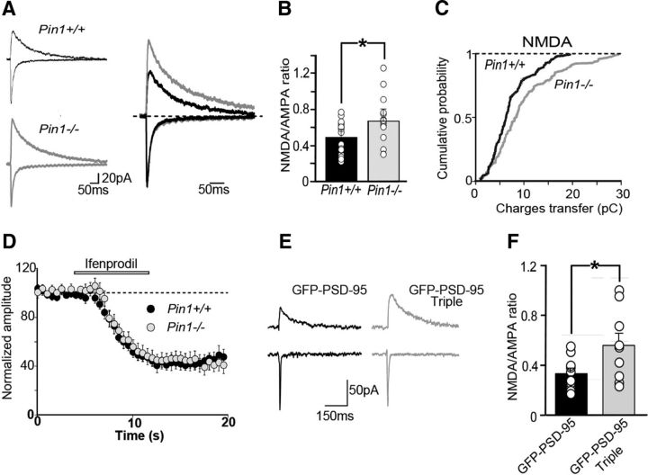
Pin1 controls synaptic signaling via NMDA receptors and regulates spine number and size. A, Sample traces of NMDAR- and AMPAR-mediated EPSCs recorded from CA1 principal cells in hippocampal slices of Pin1+/+ and Pin1−/− at holding potentials of −60 and +40 mV, respectively. Each trace is the average of 10 responses. On the right, the traces normalized to those mediated by AMPAR are superimposed. B, Summary graphs of the NMDA/AMPA-mediated receptor response ratios. Data represent mean ± SEM. Open symbols are individual values. *p < 0.05, Student's t test. C, Cumulative probability plots of charge transfers through NMDAR-mediated currents (*p = 0.003; Kolmogorov–Smirnov test). D, Time course of ifenprodil action (open bar) on NMDAR-mediated synaptic currents from Pin1+/+ (n = 12) and Pin1−/− mice (n = 10). Each point represents the mean ± SEM. E, Examples of evoked AMPA- and NMDA-mediated EPSCs in cultured hippocampal cells from GFP-PSD-95wt (black) and GFP-PSD-95 triple-mutant (gray) transfected cells. F, Summary graphs of the NMDA–AMPA ratios of transfected neurons. Open symbols are individual values. *p < 0.05, Student's t test.

