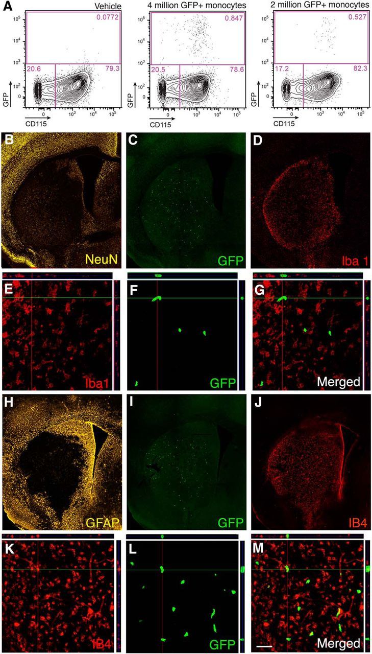
Figure 1.

Transplanted and endogenous monocytes are recruited to injured brain tissue after stroke. A, Flow cytometry analysis of blood samples from animals injected intravenously 1 d after MCAO with either vehicle (n = 2) or with 2 (n = 4) or 4 (n = 4) million GFP+ monocytes and killed 2 d later. B–G, Fluorescence microscopic images of mouse brain coronal sections showing the ischemic lesion in the striatum visualized by NeuN staining (B), distribution of grafted GFP+ monocytes within the lesion (C, F), and expression of Iba1 (D, E) by cells within the injured striatum. E–G, Confocal images showing GFP+ grafted monocytes in the lesioned striatum (F) not expressing Iba1 (E) with merged image in G. H–K, Fluorescence microscopic images of mouse brain coronal sections showing extensive GFAP staining mostly outside the lesion (H), distribution of grafted GFP+ monocytes within the lesion (I), and expression of IB4 (J, K) by cells within the injured striatum. L, M, Confocal images showing GFP+ grafted monocytes in the lesioned striatum (L) expressing activation marker IB4 (I) with merged image in M. Scale bar (in M): B–D, H–J, 420 μm; E–G, K–M, 50 μm.
