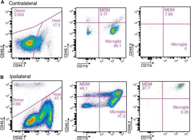
Figure 2.
Flow cytometry analysis of brain hemispheres [contralateral (A) and ipsilateral (B) to the lesion] from CD45.1 mice subjected to MCAO and injected intravenously with 4 million monocytes from CD45.2 mice on the day after the insult and killed 2 d later. Note the presence of high numbers of grafted CD45.2high/CD11bhigh and endogenous CD45.1high/CD11bhigh monocytes ipsilateral to the ischemic lesion. The CD45.1low/CD11bhigh cells are microglia.

