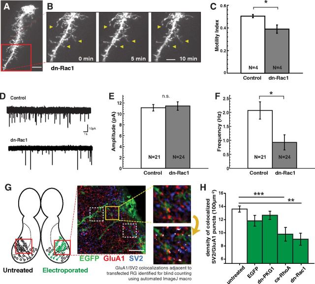
Figure 6.
Expression of dominant-negative Rac1 suppresses radial glial filopodial motility and reduces synapse density in nearby neurons. A–C, Example radial glial cell in vivo expressing dn-Rac1 is morphologically similar to a normal cell (A) but exhibits greatly reduced filopodial motility (B, C). D, Example traces of AMPA mEPSC recordings from tectal neurons surrounded by EGFPf-expressing (control) or dn-Rac1-transfected radial glia. E, F, Comparison of control neurons with neurons surrounded by dn-Rac1-expressing radial glia revealed no difference in mEPSC amplitude (E), but a profound decrease in mEPSC frequency (F). G, Synapse density was measured by counting colocalized puncta of SV2 (presynaptic) and GluA1 (postsynaptic) immunofluorescence on brain sections in tectal neuropil regions electroporated to generate dense glial expression of EGFPf alone or with constructs that reduce glial motility. Untransfected opposite hemispheres were used as control. At least six fields per animal were analyzed from 25 animals. A sample field is shown before (top) and after (bottom) automated processing to identify “anatomical synapses” (white overlay). H, Anatomical synapse density was significantly reduced in neuropil served by radial glia transfected with ca-RhoA (N = 27 fields from 4 tecta) or dn-Rac1 (N = 30 fields from 4 tecta). Neither control EGFPf (N = 45 fields from 6 tecta) nor dn-PKG1 (N = 78 fields from 11 tecta) transfection decreased anatomical synapse density compared with untransfected hemispheres (N = 159 fields from 25 tecta). *p < 0.05, Student's t test. **p < 0.01, ***p < 0.005, one-way ANOVA. Scale bars: A, G, 10 μm; B, 5 μm.

