
Figure 8.
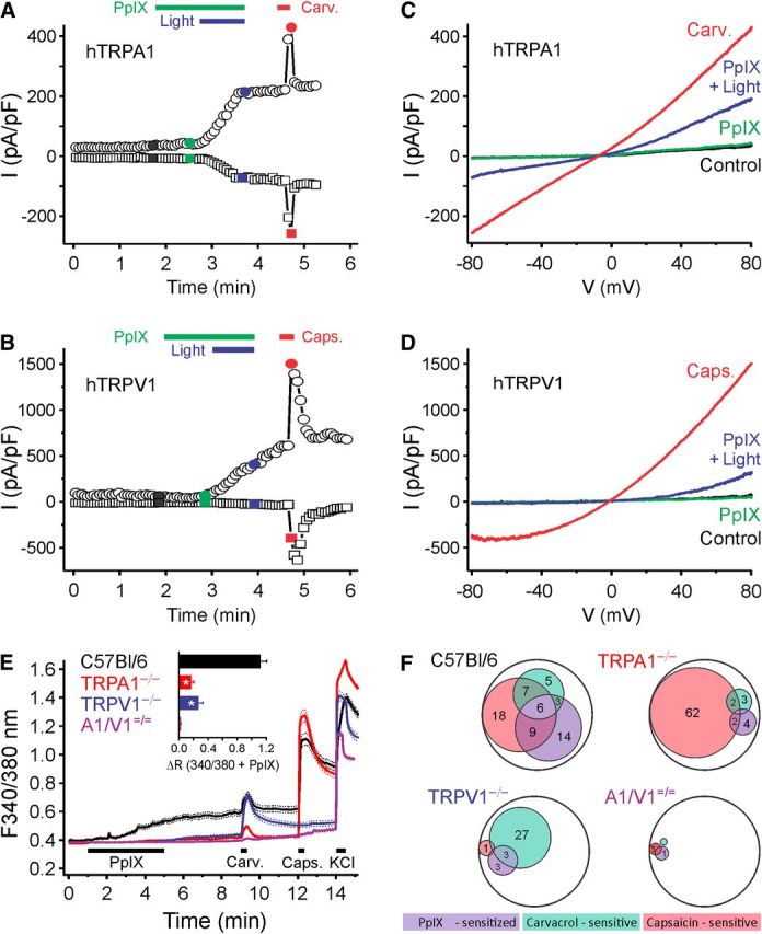
Blue light (460–490 nm) evokes hTRPA1- and hTRPV1-dependent membrane currents in the presence of PpIX. Example of a whole-cell patch-clamp recording from an (A) hTRPA1- or (B) hTRPV1-expressing HEK293t cell stimulated with PpIX (20 nm) and then blue light (60 s) in the presence of PpIX, followed by carvacrol (100 μm). Currents at −80 mV (squares) and 80 mV (circles) of voltage ramps at regular intervals were recorded. C, D, Ramp currents recorded at the times indicated by the corresponding color in A and B, respectively. E, F, PpIX activates a subpopulation of mouse DRG neurons; the effect is substantially reduced in DRGs from TRPA1−/− and TRPV1−/− mice and abolished in TRPA1−/−/TRPV1−/− mice. E, Compared with DRG neurons from WT (n = 278 cells) stimulated by PpIX (PpIX, 100 μm), the responses were substantially reduced in TRPA1−/− (n = 330), TRPV1−/− neurons (n = 209), and TRPA1−/−/TRPV1−/− (n = 266). Data are mean ± SEM. Inset, Comparison of the response amplitudes to PpIX during 340/380 illumination. *p < 0.001. F, Venn diagram of neuronal populations sensitive to PpIX, carvacrol, and capsaicin. The number reflects the respective percentage of neurons; the encompassing circle represents the total neuronal population. A–F, Carvacrol 50 μm (Carv.); and capsaicin 1 μm (Caps.).