Figure 2.

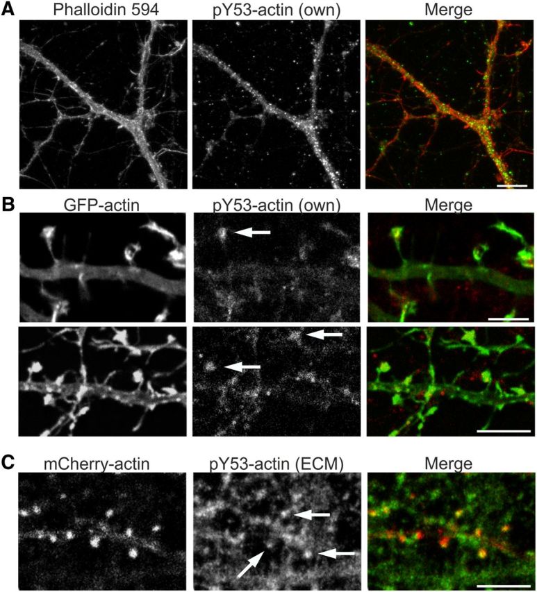
Actin Y53 phosphorylation is a strictly localized phenomenon. A, Immunofluorescence staining of primary DIV10 hippocampal low-density culture using anti-pY53-actin specific antibody. Y53-phosphorylated actin is located throughout dendrites and filopodial protrusions in a punctate manner. Scale bar, 15 μm. B, Immunofluorescence staining of primary DIV14 hippocampal neurons using pY53-actin specific antibody shows the localization of pY53-actin to dendritic spine heads. Cells are transfected with GFP-actin to highlight a single dendrite. Images from one confocal layer (z = 0.450 μm) are shown. Arrows indicate staining in spine heads. Scale bars, 5 μm. C, Immunofluorescence staining of mCherry-actin-transfected primary DIV14 hippocampal neurons using pY53-actin antibody (ECM Biosciences). Images from one confocal layer (z = 0.450 μm) are shown. Antibody staining shows clear localization in spine heads (arrows). Scale bars, 5 μm. Data in A–C represent five independent experiments.
