
Figure 5.
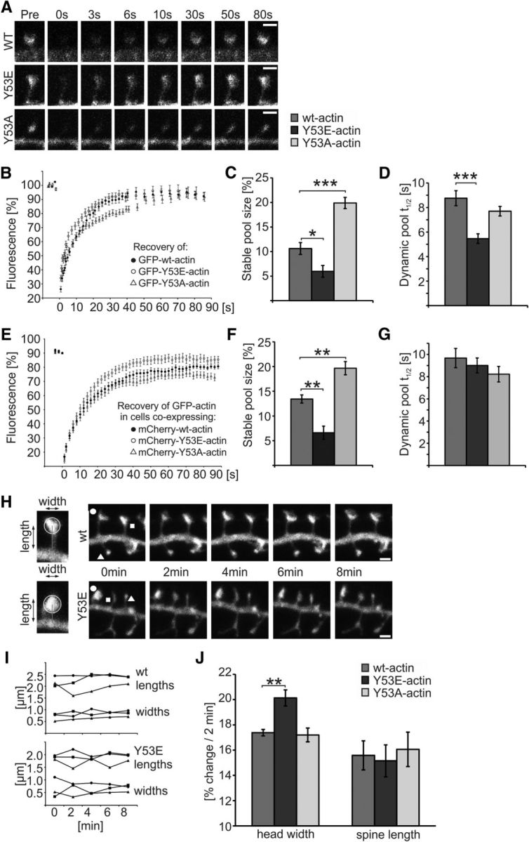
Expression of mutant actin mimicking pY53-actin increases the turnover rate of actin filaments and spine head dynamics. A, Example images presenting fluorescence recovery of GFP-(wt/Y53E/Y53A)-actin in dendritic spines of primary DIV14 hippocampal neurons. B, Analyses of FRAP assays show the mean GFP-actin fluorescence recovery curves of GFP-(wt/Y53E/Y53A)-actin. C, Stable pool sizes measured from the mean FRAP curves of individual cells show that GFP-Y53E-actin has a smaller, whereas GFP-Y53A-actin has a larger, stable-F-actin pool fraction. Stable pool size: wt = 11%, Y53E = 6%, Y53A = 20%. *p < 0.05, ***p < 0.001, one-way ANOVA with Bonferroni's post hoc test. D, GFP-Y53E-actin recovery half-time is significantly shorter, whereas GFP-Y53A-actin shows no significant change compared with GFP-wt-actin. Recovery half-time of dynamic pool: wt: 8.8 s; Y53E: 5.5 s; Y53A: 7.7 s. Data in C and D represent n(wt) = 14 cells, 51 spines, n(Y53E) = 14 cells, 50 spines, n(Y53A) = 14 cells, 55 spines pooled from three independent experiments. ***p < 0.001, one-way ANOVA with Bonferroni's post hoc test. E, Analyses of FRAP assays show the mean GFP-actin fluorescence recovery curves in dendritic spines of primary DIV14 hippocampal neurons coexpressing mCherry-tagged wt-, Y53E-, or Y53A-actin. F, Stable pool sizes measured from the mean FRAP curves of individual cells show that GFP-actin in spines coexpressing mCherry-Y53E-actin have a smaller stable-F-actin pool fraction, whereas spines coexpressing mCherry-Y53A-actin have a larger stable-F-actin pool fraction. Stable pool size: wt = 13%, Y53E = 7%, Y53A = 20%. **p < 0.01 one-way ANOVA with Bonferroni's post hoc test. G, GFP-actin recovery half-time of dynamic F-actin pool is not significantly different in cells expressing mCherry-(wt/Y53E/Y53A)-actin together with GFP-actin. Recovery half-time of dynamic pool: wt: 9.7 s; Y53E: 9.0 s; Y53A: 8.2 s. Data in F and G represent n(wt) = 15 cells, 58 spines, n(Y53E) = 14 cells, 56 spines, n(Y53A) = 15 cells, 59 spines pooled from four independent experiments. H, Representative images used in spine length and width analyses. Images of GFP fluorescence taken at 2 min intervals. Scale bars, 1 μm. I, Analysis of spine motility by measuring the mean head width and spine length fluctuation at 2 min intervals. Symbols indicating the time points refer to the spines marked with corresponding symbols in H. J, Fluctuation percentages were averaged to give a motility index for cells expressing mCherry-(wt/Y53E/Y53A)-actin together with GFP. The magnitude of spine head movement was larger in cells expressing Y53E-actin compared with wt-actin (wt: 17%, n = 6 cells, 340 spines; Y53E: 20%, n = 5 cells, 375 spines; Y53A: 17%, n = 6 cells, 420 spines). No significant change in spine length fluctuation was observed (wt: 16%, Y53E: 15%, Y53A: 16%). Data are pooled from three independent experiments. **p < 0.01 one-way ANOVA with Bonferroni's post hoc test. Data are represented as mean ± SEM.