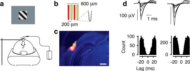
Figure 1.
Behavioral setup, recordings, and isolated single neurons. a, Setup for head-fixed behavior with air-cushioned spherical treadmill and lick-sensor. b, Schematic of the four-shank silicon probe. c, Coronal section. The four shanks of the electrode were stained in alternating fashion with DiI (yellow) and DiD (red). Blue represents DAPI. Scale bar, 400 μm. d, Average spike-waveforms and autocorrelograms of two example single units recorded from area V1. Units 144–5.x.31 and 144–4.x.5.

