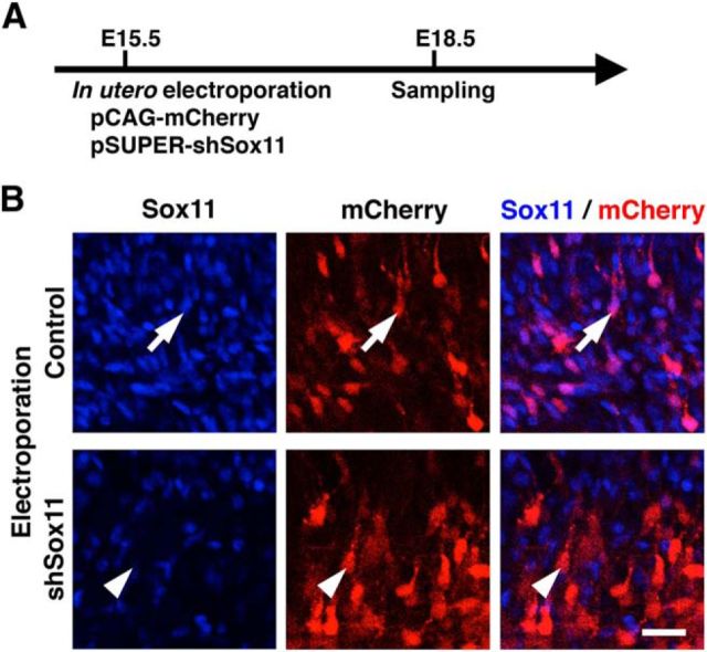
Figure 3.
Characterization of the Sox11-shRNA-expression vector. A, Experimental procedure. pCAG-mCherry (0.3 mg/ml) and pSUPER-shSox11 (1.65 mg/ml) were cotransfected into layer 2/3 neurons using in utero electroporation at E15.5, and coronal sections were prepared at E18.5 and stained with anti-Sox11 antibody and Hoechst 33342. B, Confocal microscopic images of cells transfected with either control (arrow) or shSox11 (arrowhead) vectors. mCherry-positive transfected neurons in the intermediate zone are shown. Scale bar, 25 μm.

