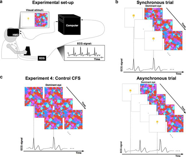
Figure 1.
Experimental setup and paradigm. a, The participant wore an HMD while her heart rate was recorded by ECG. The ECG signal was recorded in real time and was sent to a computer that generated visual stimuli flashing at the frequency of the participant's heartbeat (synchronous trials) or at a modified frequency (asynchronous trials). b, Sequence of visual stimuli presented to the participant in the synchronous (top) and asynchronous condition (bottom). The Mondrian patterns were presented to the dominant eye at a fixed frequency of 10 Hz and the target (yellow octagon) was flashed to the other eye. In synchronous trials, the flashes of the target corresponded to the moment of the QRS complex obtained from the ECG signal and reflecting systolic contraction. The task of the participants was to indicate the position of the target with respect to the fixation cross (above or below). c, CFS control experiment (Experiment 4) in which Mondrian patterns and the target stimuli were presented to both eyes without any binocular rivalry to control for detection and response biases.

