Figure 4.
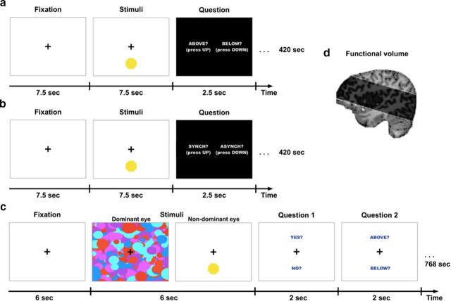
High-resolution imaging paradigms. a, fMRI localization task in which participants viewed the same stimuli used in Experiments 1 and 2 but with no binocular masking. Participants were asked to report the location (above or below fixation) of the octagon which, unbeknownst to them, was flashing synchronously or asynchronously with their heartbeat. b, fMRI heartbeat awareness task in which participants viewed the exact same stimuli as the localization task but were now informed that the flashing was related to their heartbeat and were requested to detect whether the flashes were synchronous or asynchronous to their current heartbeat. This was used as a functional localizer for the anterior insula regions. c, CFS localization task. In Experiment 9, the octagon (flashing synchronously or asynchronously to their heartbeat) was rendered continuously invisible by high-contrast masks presented to the dominant eye (as in Experiments 1 and 2). Participants were asked to guess the location of the stimuli and report whether they saw the target at any time during the trial. d, Functional volume scanned in high-resolution fMRI at 7T.

