
Figure 4.
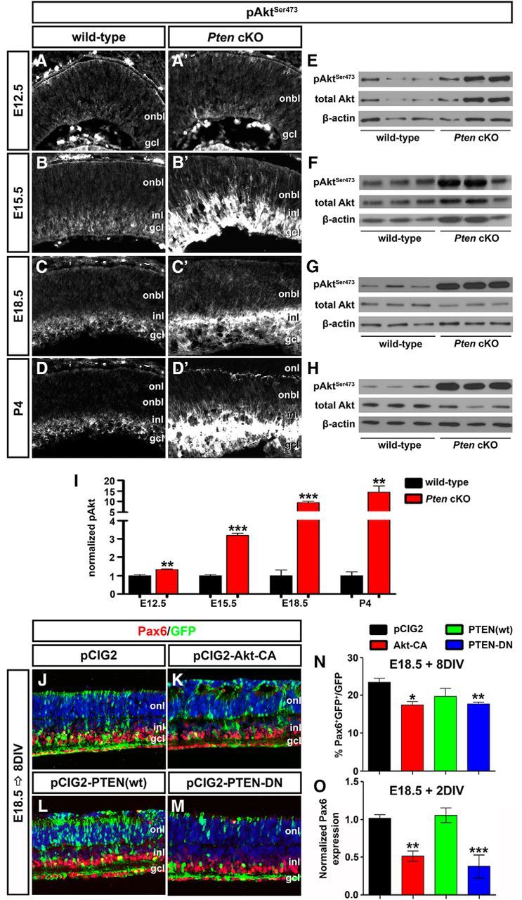
Hyperactivated Akt in Pten cKO retinas contributes to the decline in amacrine cell differentiation. A–D′, Immunolabeling of wild-type and Pten cKO retinas at E12.5 (A, A′), E15.5 (B, B′), E18.5 (C, C′), and P4 (D, D′) for pAktSer473. E–I, Western blot analysis and densitometry of pAktSer473 in wild-type and Pten cKO retinal lysates at E12.5 (E), E15.5 (F), E18.5 (G), and P4 (H). Levels of pAktSer473 are elevated at all stages analyzed when Pten is deleted (I). J–M, E18.5 retinas electroporated with pCIG2 control (J), Akt-CA (K), PTEN(wt) (L), or PTEN-DN (M) and cultured for 8 DIV. GFP+ (green) electroporated amacrine cells were identified by Pax6 immunolabeling (red). Blue is a DAPI counterstain. N, Percentages of GFP+ amacrine cells (GFP+Pax6+) after electroporation of pCIG2, Akt-CA, PTEN(wt), or PTEN-DN. O, qPCR to assess Pax6 transcript levels in GFP+ cells sorted by FACS after electroporation of pCIG2, Akt-CA, PTEN(wt), or PTEN-DN. *p < 0.05; **p < 0.01; ***p < 0.001.