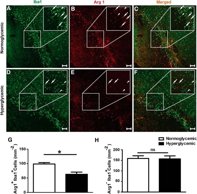
Figure 3.
Hyperglycemia reduces the number of Arg1 positive noninflammatory myeloid cells in the peri-infarct zone 48 h after MCAO. A–F, Representative confocal images of Iba1 (green, marker of microglia and monocytes/macrophages) and Arg1 (red) staining in normoglycemic and hyperglycemic mice. White arrows, Arg1+Iba+ cells; white arrowheads, Arg1−Iba+ cells. G, H, Quantification of Arg1+Iba+ and Arg1−Iba+ cells per square millimeter. Unpaired t test, **p < 0.003 (n = 6–8 mice/group) .Scale bar 100 μm. Data are shown as means ± SEM.

