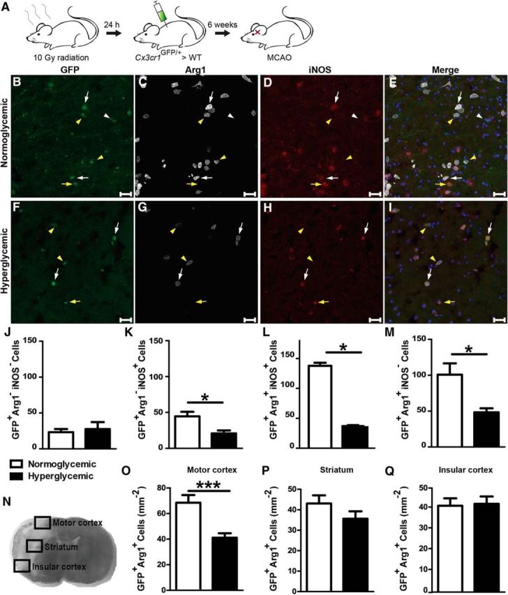
Figure 4.

Acute hyperglycemia during MCAO modified the polarization of infiltrating monocytes/macrophages. A, Infiltrating monocytes/macrophages were labeled by transplantation of Cx3cr1GFP/+ bone marrow. B–I, Forty-eight hours after MCAO, Cx3cr1GFP/+ cells (green) were stained for Arg1 (white) and iNOS (red). Cx3cr1GFP/+ cells in the peri-infarct area of the motor cortex were Arg1−iNOS− (white arrowheads), Arg1−iNOS+ (yellow arrows, inflammatory macrophages), Arg1+iNOS+ (white arrows), or Arg1+iNOS− (yellow arrowheads, noninflammatory macrophages). Scale bar, 50 μm. J–M, Quantification of the four cell types in coronal sections of the whole brain revealed a lower density of inflammatory macrophages, but mainly the Arg1+iNOS+ and noninflammatory macrophages were reduced. N, To localize the hyperglycemic effect on noninflammatory polarization in the peri-infarct tissue, three distinct areas where analyzed separately. O–Q, Cx3cr1GFP/+Arg1+ cells in the targeted regions revealed a significant difference in the motor cortex, whereas there was no change in the striatum or insular cortex. Unpaired t test, *p < 0.05, ***p < 0.0005, data are shown as means ± SEM (n = 4/group for whole section and n = 10 mice/group for targeted regions).
