Figure 7.
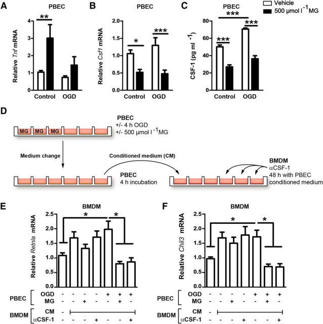
By reducing expression and release of CSF-1 in PBECs, MG indirectly impairs noninflammatory polarization of macrophages. A, MG treatment increased Tnf expression in PBECs under control conditions and after OGD. Two-way ANOVA, F(1, 65) = 8.58, p = 0.0047. *p < 0.05, **p < 0.01, ***p < 0.001 (n = 18 with 6 wells in 3 independent experiments, Bonferroni posttests). B, C, In contrast, MG reduced mRNA levels of Csf1 (F(1, 68) = 23.78, p < 0.0001) and the release of CSF-1 from PBECs (F(1, 16) = 134.7, p < 0.0001, n = 18 with 6 wells in 3 independent experiments). D, To investigate the crosstalk of PBEC with BMDM by the secretion of CSF-1, BMDMs were treated with the conditioned medium (CM) of PBECs that were stimulated with MG and OGD as indicated. E, F, In BMDMs, the expression of Retnla and Chil3, typical marker genes of noninflammatory macrophages, was diminished by the CM of MG- and OGD-challenged PBECs. A comparable effect was obtained by adding CSF-1-neutralizing antibodies (α-CSF-1) to CM. Expression was normalized to controls that did not receive CM. One-way ANOVA, *p < 0.05, **p < 0.01, ***p < 0.005 (n = 15–28, Tukey's multiple-comparisons test). Data are shown as means ± SEM.

