Figure 2.

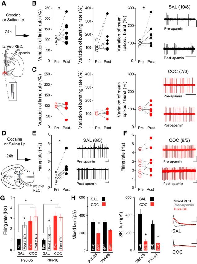
SK channels contribute to increased burst firing of VTA DA neurons following cocaine (COC). A, Schematic of the in vivo experimental design using double-barrel pipette for microinjection. B, In saline (SAL)-treated rats, application of apamin increased the firing rate (left) and spikes per burst (right) of VTA DA neurons. No effect is observed on the bursting rate (middle). Representative traces from SAL-treated rats. Scale bars: horizontal, 1 s; vertical, 1 mV. C, Application of apamin had no effect on firing rate, burst firing, or spikes per burst in COC-treated rats. Representative traces from COC-treated rats. Scale bars: horizontal, 1 s; vertical, 1 mV. D, Schematic of ex vivo recording experiments. E, F, In cell-attached recordings, apamin application increased the firing rate of VTA DA neurons in SAL-treated mice, but had no effect in mice receiving COC. Inset, Representative traces. Scale bars: horizontal, 1 s; vertical, 20 mV. G, In both adolescent (P28–P35) and adult (P84–P98) mice, basal firing rate was increased in COC-treated relative to SAL-treated mice, and apamin application increased the firing rate in SAL-treated mice only. H, The mixed and SK-mediated components of the IAHP were reduced following COC treatment in both adolescent and adult mice. Right, Representative traces. Scale bars: horizontal, 50 ms; vertical, 50 pA. *p < 0.05.
