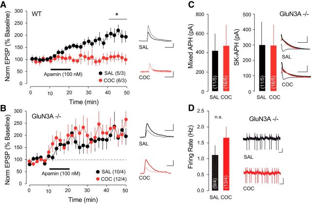
Figure 3.
SK channel function in VTA DA neurons in GluN3A−/− mice is preserved after cocaine (COC). A, In VTA DA neurons, bath application of apamin potentiated NMDA-mediated EPSP amplitude, which was occluded 24 h after COC treatment. Inset, Representative traces showing averaged EPSP 5 min immediately before apamin application and 20 min after application. Scale bars: horizontal, 50 ms; vertical, 1 mV. B, COC treatment did not occlude apamin-induced LTP of NMDA-mediated EPSPs in GluN3A−/− mice. Inset, Representative traces showing averaged EPSP 5 min immediately before apamin application and 20 min after application. Scale bars: horizontal, 50 ms; vertical, 1 mV. C, The mixed and SK-mediated components of the IAHP were not reduced by COC treatment in GluN3A−/− mice. Inset, Representative traces. Scale bars: horizontal, 100 ms; vertical, 50 pA. D, COC-induced increase in firing rate of VTA DA neurons was not significant in GRIN3A knock-out mice. Scale bars: horizontal, 1 s; vertical, 20 mV. *p < 0.05. SAL, Saline.

