Figure 6.
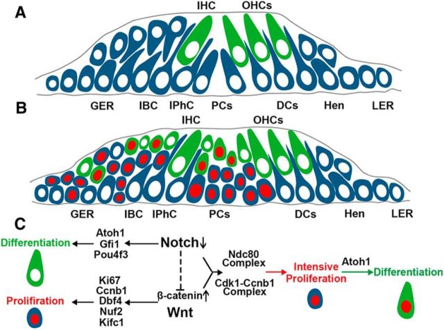
Model of molecular mechanism during HC generation. A, Schematic cross-section of the organ of Corti of a neonatal mouse cochlea. B, Schematic cross-section of the organ of Corti of a neonatal mouse cochlea during HC generation. Blue cells indicate the Sox2+ cells, gray cells indicate Lgr5+ SCs, green cells indicate Myo7a+ HCs, and red nuclei indicate the EdU+ cells. C, Activation of Notch signaling inhibits the differentiation of HCs by inhibiting Atoh1, Gfi1, and Pou4f3. Notch signaling serves as a negative regulator for the proliferation of progenitors through inhibiting Wnt signaling. The activation of Wnt signaling promotes the proliferation of SCs through upregulating Ki67, Ccnb1, Dbf4, Nuf2, and Kifc1. In β-cat-OE/Notch1-KO mice, more cell-cycle-related genes are upregulated and lead to intensive proliferation of SCs, including the Ndc80 complex and Cdk1–cnb1. The proliferated SCs could still react to Atoh1 and differentiate into HCs. OHC, Outer HC; IHC, inner HC; IBC, inner border cell; IPhC, inner phalangeal cell; PC, pillar cell; DCs, Deiters' cells; Hen, Hensen's cells; LER, lesser epithelial ridge.

