Figure 7.

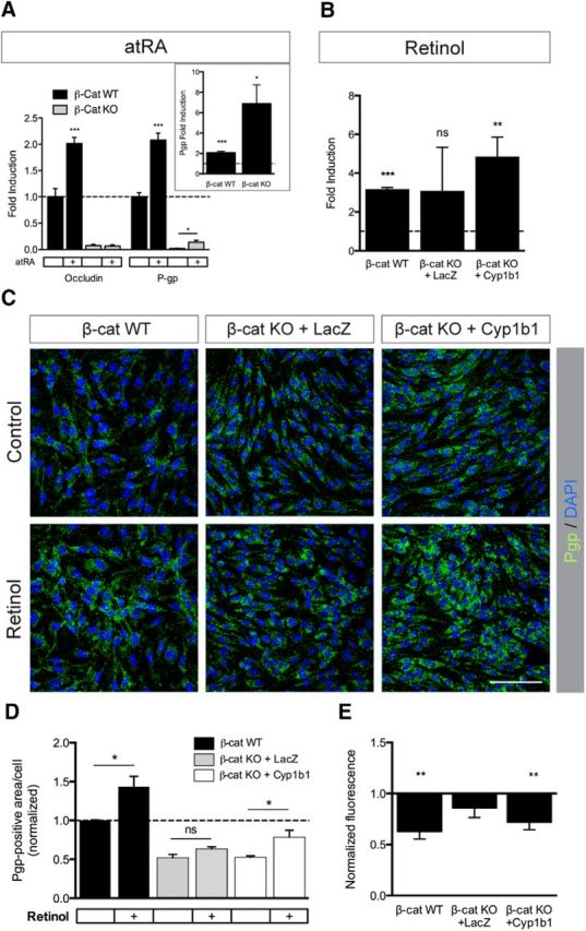
Cyp1b1-mediated metabolism of retinol increases Pgp expression and activity. A, qRT-PCR analysis for Ocln and Pgp in β-catenin WT and β-catenin KO ECs. Cells were treated with 5 μm atRA for 48 h. Expression levels are shown relative to control (dashed line, n = 5). Inlay represents Pgp expression in β-catenin WT and β-catenin KO ECs after atRA treatment. B, qRT-PCR for Pgp in β-catenin WT, β-catenin KO infected with LacZ as control and β-catenin KO ECs reinfected with Cyp1b1. ECs were treated with 5 μm retinol for 72 h. Expression levels under control conditions were defined as 1 (n = 4). C, Representative pictures of immunofluorescence stainings for Pgp on β-catenin WT, β-catenin KO infected with LacZ and β-catenin KO ECs reinfected with Cyp1b1. Treatment was the same as for B (n = 3, 10 pictures/condition). Scale bar, 100 μm. D, Quantification of Pgp-positive area per cell relative to β-catenin WT ECs under control condition based on IF staining. E, Rhodamine-123 efflux assay of β-catenin WT, LacZ- and Cyp1b1-infected β-catenin KO ECs treated with 5 μm retinol for 72 h (n = 5). Bars represent normalized fluorescence relative to untreated. *p < 0.05. **p < 0.01. ***p < 0.001. ns, Not significant. Error bars indicate SEM.
