Figure 6.

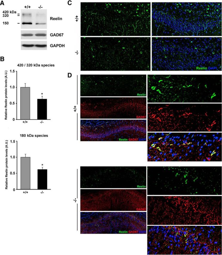
Reduced expression of Reelin in the hippocampus and stratum oriens of Ndel1 CKO mice. A, Expression of all Reelin species (180, 320, 420 kDa) in the hippocampus of 10-week-old Ndel1 CKO mice as detected by Western blots. Note the unchanged levels of the interneuronal marker GAD67 in the mutant animals compared with WT littermates. B, Bar graphs for the arbitrary levels of Reelin in the hippocampus of 10-week-old animals. 420/320 kDa species for WT (+/+): 1.00 ± 0.12 versus Ndel1 CKO (−/−): 0.64 ± 0.09; mean ± SE (n = 3 animals). 150 kDa species for WT (+/+): 1.00 +/− 0.09 versus Ndel1 CKO (−/−): 0.62 ± 0.07; mean ± SE (n = 3 animals), *p < 0.05 by Student's t test. C, Representative confocal images from a 7-week-old Ndel1 CKO mouse and WT littermate depicting the reduced number of Reelin-positive cell bodies in stratum oriens and reduced immunoreactivity for Reelin in CA1. D, Representative confocal images of the CA1 region from a 7-week-old Ndel1 CKO mouse and WT littermate confirming the reduced levels of Reelin in GAD67-positive interneurons of stratum oriens above the CA1 subfield.
