Figure 5.

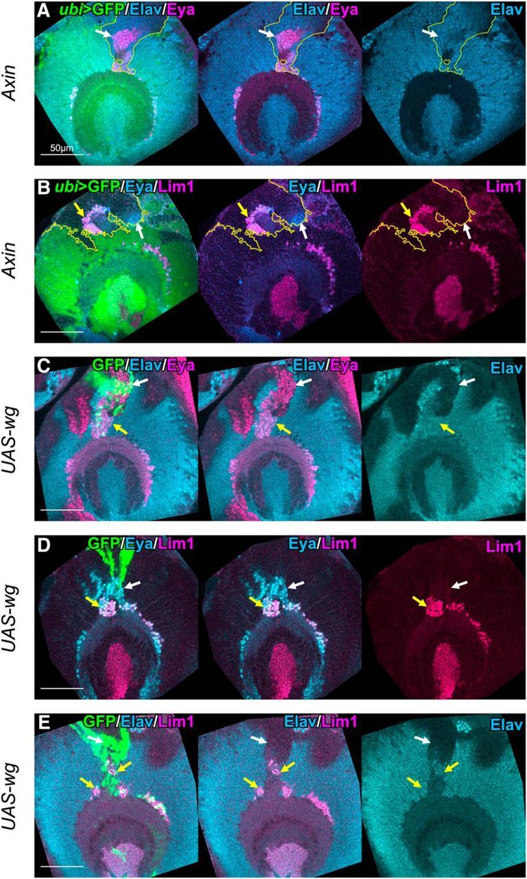
Wnt signaling induces ectopic GPC-type neurons in the anterior region of OPC. Clones generated in the anterior region of OPC were examined. A, B, Axin mutant clones are labeled by the absence of GFP signals (green). A, Eya+ (magenta)/Elav+ (blue) neurons were ectopically observed in the clones (white arrows). B, Lim1+ (magenta)/Eya+ (blue) cells (yellow arrows) and Lim1−/Eya+ cells (white arrows) appeared ectopically in the clones. C–E, Clones expressing wg are labeled by GFP (green). C, Eya+ (magenta)/Elav+ (blue) neurons were ectopically observed around the clones (yellow arrows). D, Lim1+ (magenta)/Eya+ (blue) cells (yellow arrows) and Lim1−/Eya+ cells (white arrows) appeared ectopically around the clones. E, Lim1+ (magenta)/Elav+ (blue) neurons were observed ectopically around the clones (yellow arrows). C, E, Neuronal differentiation was occasionally suppressed, as indicated by white arrows.
