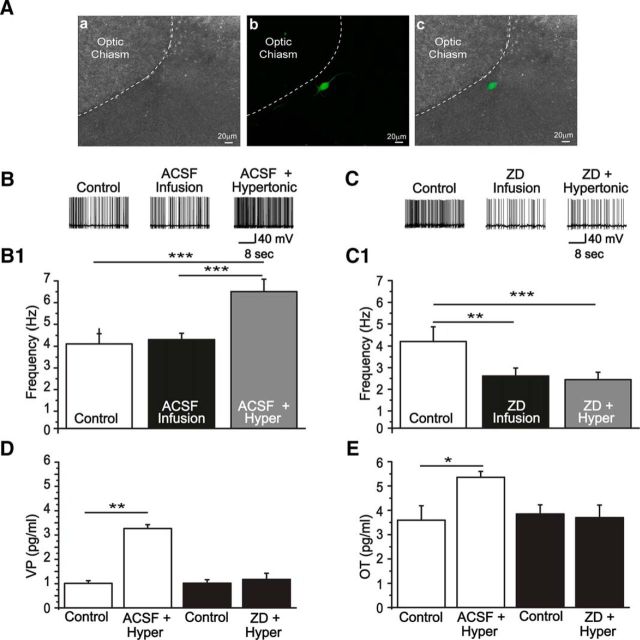
Figure 9.
Physiological implications of HCN channel blockade in the SON. A, Immunofluorescence reveals the anatomical localization of neurons recorded using the in situ brainstem-hypothalamic preparation: a, Bright-field; b, biocytin-labeled neuron reveled by streptavidin; c, a and b merged images. B, C, Representative traces of two magnocellular neurons recorded in a control condition, after ACSF (B) or ZD7288 infusion (C), and hypertonic stimulation (Hyper). B1, Bar graphs are average firing frequencies of neurons recorded during the control, after isotonic ACSF infusion and hypertonic stimulus (n = 5 rats). ** p < 0.005 (ANOVA One Way followed by Bonferroni post hoc test). *** p < 0.001 (ANOVA One Way followed by Bonferroni post hoc test). ACSF infusion did not change the firing rate of MNCs, and an increase is observed during hypertonic stimulation. C1, Average firing frequencies of neurons in the control condition, after ZD7288 infusion followed by hypertonic stimulation. In this case, previous blockade of HCN channels avoided the increment in firing rate induced by hypertonicity. D, E, Averaged concentrations of vasopressin and oxytocin measured in the effusate, using radioimmunoassay, for each situation. ZD7288 (E) prevented the increase in the concentration of VP and OT induced by hypertonicity (D). *p < 0.05 (paired Student's t test). **p < 0.005 (paired Student's t test).

