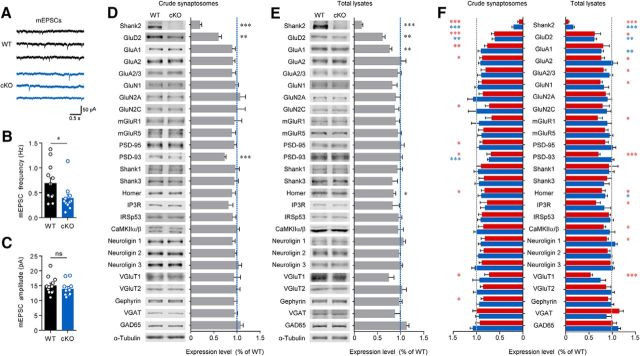
Figure 6.
Reduced mEPSC frequency and synaptic levels of GluD2 and PSD-93 in the cerebellum of Pcp2-Cre;Shank2fl/fl mice. A–C, Reduced frequency but normal amplitude of mEPSCs in Pcp2-Cre;Shank2fl/fl (cKO) PCs (P19–P23). n = 16 cells from 7 (3 male, 4 female) mice for WT and 16 cells from 6 (3 male, 3 female) mice for cKO. *p < 0.05 (Student's t test). D, E, Reduced synaptic levels of GluD2 and PSD-93 in the Pcp2-Cre;Shank2fl/fl cerebellum (P21–P22), as determined by immunoblotting of crude synaptosomes (D). The total level of GluD2, but not PSD-93, was reduced, as determined by immunoblotting of total lysates (E). n = 6 (2 male, 4 female) for WT and cKO. *p < 0.05 (Student's t test). **p < 0.01 (Student's t test). ***p < 0.001 (Student's t test). F, Side-by-side comparison of synaptic protein levels in crude synaptosomes and total lysates from Shank2−/− (red) and Pcp2-Cre;Shank2fl/fl (blue) mice. The data presented in Figures 3A, B and 6D, E were reused here for comparison.

